Guided Surgery for Dental Implants: A Comprehensive Overview
Guided implant surgery, utilizing advanced computer programs, enables precise tilted dental implant placement, even in dense bone, revolutionizing the procedure.
Custom surgical guides, embodying prosthodontic planning, streamline workflows, while flapless techniques minimize invasiveness for single tooth replacement.
Immediate loading protocols demonstrate effectiveness and predictability, and single drill techniques reduce bleeding and time, even for patients with coagulopathy.
Guided implant surgery represents a significant evolution in restorative dentistry, merging advanced imaging, digital planning, and precise surgical execution. Traditionally, implant placement relied heavily on the surgeon’s experience and visual assessment. However, the advent of computer-aided implant surgery allows for a more predictable and accurate approach.
This methodology utilizes surgical guides – physical templates fabricated from digital models – to direct drill placement and implant insertion. These guides are based on Cone Beam Computed Tomography (CBCT) scans and digital impressions, offering a detailed three-dimensional representation of the patient’s anatomy.
The application of this technology extends to complex cases, including those requiring tilted implants or flapless surgery, ultimately enhancing treatment outcomes and patient satisfaction. It’s a collaborative process involving surgeons, prosthodontists, and dental technicians.
What are Dental Implants?
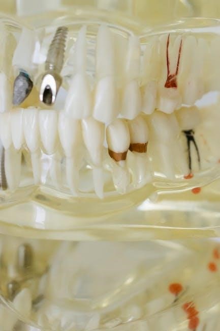
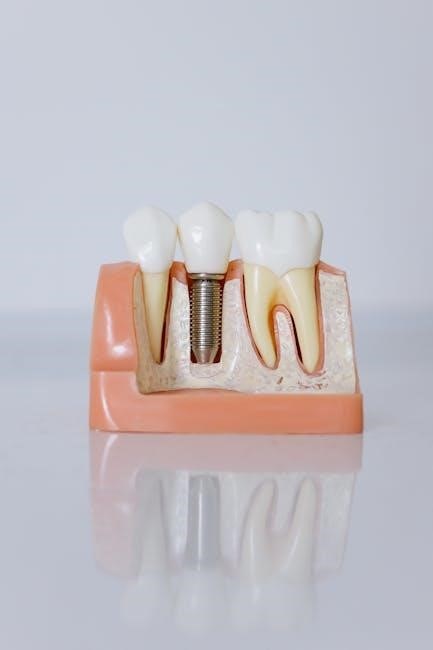
Dental implants are artificial tooth roots, typically made of biocompatible titanium, surgically placed into the jawbone to support a replacement tooth or bridge. They offer a robust and long-lasting solution for missing teeth, functioning much like natural teeth and preventing bone loss. Unlike dentures, implants are anchored directly to the jaw, providing superior stability and comfort.
The process involves osseointegration, where the implant fuses with the surrounding bone, creating a strong foundation. Once integrated, a final restoration – a crown, bridge, or denture – is attached to the implant via an abutment.
Guided surgery enhances implant placement accuracy, ensuring optimal positioning for successful osseointegration and long-term functionality, even in challenging cases where bone density is a concern.
The Evolution of Implant Dentistry
Implant dentistry has dramatically evolved from early, less predictable techniques. Initially, implant placement relied heavily on the surgeon’s experience and visual assessment. The introduction of cone beam computed tomography (CBCT) marked a significant leap, providing 3D imaging for improved treatment planning.
However, the true revolution arrived with computer-aided design and manufacturing (CAD/CAM) technology, enabling guided surgery. This allows for virtual implant placement and the creation of precise surgical guides, minimizing invasiveness and maximizing accuracy.
Today, advancements continue with techniques like immediate loading and flapless surgery, further streamlining the process and enhancing patient outcomes, all stemming from the initial pursuit of more predictable and reliable implant procedures.

The Benefits of Guided Surgery
Guided surgery offers increased precision, reduced surgical risks, minimized discomfort, and shorter treatment times, improving predictability and patient experience significantly.
Increased Precision and Accuracy
Guided surgery dramatically elevates the precision and accuracy of dental implant placement. Utilizing pre-operative CBCT scans and digital planning, surgeons can visualize the ideal implant position and angulation before even making an incision.
This meticulous planning translates into a surgical guide that directs the drill, ensuring adherence to the pre-determined plan. The application of computer programs allows for tilted implant placement, maximizing bone utilization, particularly in areas of limited space.
This level of accuracy minimizes deviations from the intended surgical plan, leading to improved prosthetic outcomes and a more predictable, reliable restoration. The summarized concept of all parties is embodied in the surgical guide.
Reduced Surgical Risks
Guided surgery significantly reduces several surgical risks associated with traditional dental implant procedures. By pre-planning the implant placement, the risk of damaging vital anatomical structures – such as nerves and sinuses – is substantially minimized.
The use of a surgical guide limits the depth and angulation of the drill, preventing accidental perforation or injury. Furthermore, techniques like flapless surgery, often facilitated by guided approaches, reduce trauma to the surrounding tissues, leading to less bleeding and swelling.
Even patients with conditions like coagulopathy can benefit, as single drill techniques reduce operative time and bleeding. This translates to a safer and more predictable surgical experience for the patient.
Minimized Post-Operative Discomfort
Guided surgery contributes to a significantly more comfortable post-operative experience for patients undergoing dental implant treatment. The precision offered by surgical guides and pre-planned procedures minimizes trauma to the surrounding soft tissues and bone.
Techniques like flapless surgery, frequently employed with guided approaches, eliminate the need for extensive incisions, resulting in less swelling, bruising, and pain. Reduced surgical trauma also translates to decreased reliance on post-operative pain medication.
Furthermore, accurate implant placement minimizes the risk of post-operative complications, such as nerve irritation or sinus issues, which can contribute to prolonged discomfort. This leads to faster healing and a quicker return to normal function.
Shorter Treatment Times
Guided surgery streamlines the dental implant process, leading to demonstrably shorter overall treatment times for patients. The pre-operative planning phase, utilizing CBCT scans and digital models, minimizes chair-side time and reduces the need for adjustments during surgery.
The use of precise surgical guides allows for accurate implant placement on the first attempt, avoiding potential complications and the need for additional procedures. Techniques like single-drill protocols further expedite the surgical phase, reducing procedure duration.
Moreover, immediate loading protocols, often facilitated by guided surgery, enable the placement of temporary restorations on the implants immediately after surgery, eliminating the waiting period for osseointegration in suitable cases.
The Guided Surgery Process: Step-by-Step
Guided surgery involves consultation, CBCT scanning, digital impressions, virtual planning, surgical guide design and fabrication, and finally, precise implant placement.
Initial Consultation and Treatment Planning
The initial consultation is paramount, establishing patient needs and expectations. A thorough clinical examination, alongside a detailed review of the patient’s medical history, forms the foundation. This phase involves discussing treatment options, including the benefits of guided surgery versus traditional methods.
Treatment planning then commences, focusing on achieving optimal functional and aesthetic outcomes. This requires a collaborative approach, often involving a surgeon and restorative dentist. The surgical guide carries all the information of prosthodontically guided implant planning, summarizing the concept of all parties involved. Careful consideration is given to bone volume, soft tissue health, and the desired final restoration.
Verification of the surgical guide fit is crucial, ensuring complete seating on a model or directly in the patient’s mouth before the surgical phase.
Cone Beam Computed Tomography (CBCT) Scanning
CBCT scanning is a cornerstone of guided implant surgery, providing a three-dimensional radiographic image of the patient’s jawbone. This detailed imaging allows for precise assessment of bone density, nerve location, and sinus proximity – critical factors for safe and effective implant placement.
Unlike conventional two-dimensional X-rays, CBCT offers a comprehensive view, enabling surgeons to visualize anatomical structures with exceptional clarity. This minimizes the risk of damaging vital nerves or anatomical landmarks during the surgical procedure. The scan data is then digitally imported into specialized planning software.
The accuracy of guided surgery relies heavily on the quality of the CBCT scan, ensuring precise virtual implant placement.
Digital Impression and Model Creation
Following the CBCT scan, a precise digital impression of the patient’s mouth is captured. This eliminates the need for messy traditional impressions using alginate or putty materials. Intraoral scanners create a highly accurate digital replica of the dentition and surrounding soft tissues.
This digital data is then used to generate a virtual model of the patient’s jaw. This model serves as the foundation for virtual implant planning and surgical guide design. The digital workflow ensures exceptional accuracy and eliminates potential distortions associated with conventional model creation.
Surgical guides are designed based on this digital model, ensuring a perfect fit and precise implant placement.
Virtual Implant Placement and Surgical Guide Design
Utilizing specialized software, the dentist virtually places implants on the digital model, considering anatomical structures and desired prosthetic outcomes. This allows for optimal implant positioning, angulation, and depth before any surgical intervention. The software facilitates planning for tilted implants when insufficient bone volume exists.
Once implant positions are determined, the software designs a surgical guide tailored to the patient’s anatomy. This guide incorporates drill sleeves that dictate the precise path and depth for each implant.
The surgical guide embodies the prosthodontically guided implant planning, summarizing the collaborative concept of all involved parties.
Surgical Guide Fabrication
Following the virtual design, the surgical guide is fabricated using various technologies. Typically, this involves 3D printing or milling from materials like acrylic resins, PEEK, or titanium. Ensuring a precise fit is crucial; the guide must seat completely and securely on the patient’s dentition or mucosa.
Before surgery, verification of the guide’s fit is performed on the model and, ideally, intraorally. Complete seating guarantees accurate transfer of the virtual plan to the surgical site.
A well-fabricated guide carries all the information of the prosthodontically guided implant planning, representing a summarized concept of all parties involved.

Types of Surgical Guides
Surgical guides are categorized as static or dynamic, and can be mucosa-supported or bone-supported, each offering unique advantages for implant placement.
Static Surgical Guides

Static surgical guides represent the most commonly utilized type in guided implant surgery. These guides are fabricated pre-operatively based on detailed CBCT scans and digital impressions, providing a fixed pathway for drill placement.
Essentially, they are passive guides, meaning they don’t actively adjust during the surgical procedure. They rely on precise fit and seating to ensure accurate implant angulation and depth. The guide carries all the information of prosthodontically guided implant planning, summarizing the collaborative concept.
Verification of the guide’s fit, both on a model and intraorally, is crucial before surgery. They are particularly effective in straightforward cases and contribute to increased precision and reduced surgical risks.
Dynamic Surgical Guides
Dynamic surgical guides represent a more advanced approach to guided implant surgery, offering adaptability during the procedure. Unlike static guides, these guides allow for adjustments based on intraoperative findings, providing flexibility when encountering unforeseen anatomical variations.
They often incorporate features like sleeves that can be repositioned or adjusted, allowing the surgeon to modify the implant trajectory if necessary. This is particularly useful in cases with limited interocclusal space or complex anatomical structures.
While more complex to design and fabricate, dynamic guides can enhance precision and predictability, especially when dealing with challenging clinical scenarios, offering a refined level of control.
Mucosa-Supported vs. Bone-Supported Guides
Surgical guides are broadly categorized as either mucosa-supported or bone-supported, each offering distinct advantages depending on the clinical situation. Mucosa-supported guides rest on the soft tissues, providing guidance for initial osteotomies but requiring careful stabilization.
Bone-supported guides, conversely, are designed to fit directly onto the underlying bone, offering enhanced stability and accuracy, particularly crucial in areas with limited soft tissue. These guides are often preferred for complex cases or when precise implant placement is paramount.
The choice between the two depends on factors like bone volume, soft tissue quality, and the surgeon’s preference, impacting the overall predictability of the guided surgery process.
Advanced Techniques in Guided Surgery
Advanced methods include flapless procedures, immediate loading, tilted implant placement, and utilizing single drill techniques for reduced invasiveness and efficiency.
Flapless Implant Surgery
Flapless implant surgery represents a significant advancement within guided surgery, offering a minimally invasive approach to tooth replacement. This technique, facilitated by precisely fabricated surgical guides, eliminates the need for traditional gum incisions and flap reflection.
Instead, access is gained directly through the gingiva, minimizing trauma to surrounding tissues. This results in reduced post-operative discomfort, swelling, and bleeding for the patient. The use of custom surgical guides ensures accurate implant placement, even in challenging anatomical situations.
Flapless procedures are particularly well-suited for single tooth replacements and cases with sufficient bone volume. It promotes faster healing times and improved aesthetic outcomes, making it a popular choice for both clinicians and patients seeking a conservative treatment option.
Immediate Loading Protocols
Immediate loading protocols, increasingly integrated with guided surgery, represent a paradigm shift in dental implant treatment. This approach involves placing a functional restoration – a crown, bridge, or denture – onto newly placed implants immediately or shortly after surgery.
Extensive research consistently demonstrates the effectiveness and predictability of immediate loading when carefully selected criteria are met. Factors like implant stability, bone quality, and occlusal forces are crucial for success. Guided surgery plays a vital role by ensuring precise implant positioning, maximizing primary stability.
Benefits include reduced treatment time, improved patient satisfaction, and preservation of soft tissue contours. However, meticulous treatment planning and adherence to established protocols are essential for optimal outcomes.
Tilted Implant Placement
Tilted implant placement, facilitated by guided surgery, offers a compelling solution for patients with limited bone volume in the posterior mandible or maxilla. This technique strategically angles implants to maximize bone contact and avoid critical anatomical structures like the inferior alveolar nerve.
Utilizing computer-aided planning and custom surgical guides, clinicians can precisely determine the optimal implant angulation. This approach often eliminates the need for extensive bone grafting procedures, reducing treatment complexity and healing times. The application of guided implant surgery allows for placement in dense cortical bone.
Tilted implants provide enhanced prosthetic support and improved esthetics, particularly when restoring edentulous arches. Careful case selection and meticulous execution are paramount for long-term success.
Use of Single Drill Techniques
Single drill techniques, increasingly integrated with guided surgery, represent a significant advancement in implant dentistry, streamlining the osteotomy preparation process. Traditionally, multiple drill steps were required to gradually expand the bone to the desired implant diameter.
However, with precise surgical guides and advanced drill designs, a single drill can often achieve the final implant site dimensions, minimizing trauma to the surrounding tissues. A recent case report demonstrated that this method reduced bleeding and overall procedure time, even in a patient presenting with coagulopathy.
This simplified approach enhances predictability and efficiency, contributing to a more comfortable patient experience and potentially faster healing.
Materials Used in Surgical Guide Fabrication
Surgical guides are commonly fabricated from acrylic resins, polyetheretherketone (PEEK), or titanium, each offering unique properties for precision and biocompatibility.
Acrylic Resins
Acrylic resins represent a frequently utilized material in the fabrication of surgical guides due to their cost-effectiveness and ease of processing. These materials allow for relatively straightforward design and manufacturing, making them accessible for many dental laboratories. However, it’s crucial to acknowledge that acrylic resins exhibit a degree of polymerization shrinkage during processing, potentially impacting the dimensional accuracy of the final guide.
Despite this limitation, advancements in acrylic resin formulations have improved stability and reduced shrinkage. They are generally suitable for static guides where high precision is needed, but the material’s mechanical properties might not be ideal for dynamic guides experiencing significant forces. Careful consideration of the specific clinical application is essential when selecting acrylic resins for surgical guide production.
Polyetheretherketone (PEEK)
Polyetheretherketone (PEEK) is gaining prominence as a material for surgical guide fabrication, offering several advantages over traditional acrylic resins. Notably, PEEK demonstrates superior mechanical strength, thermal stability, and biocompatibility. Its radiolucency is also beneficial, allowing for clear visualization of the implant site during CBCT scans and intraoperative assessment.
Unlike acrylics, PEEK exhibits minimal polymerization shrinkage, contributing to enhanced dimensional accuracy and fit of the surgical guide. While generally more expensive than acrylic resins, the improved properties of PEEK justify the cost for complex cases or when greater precision and durability are required. It’s suitable for both static and, with appropriate design, dynamic surgical guides.
Titanium
Titanium, a biocompatible and robust metal, represents a premium material choice for fabricating surgical guides, particularly for demanding clinical scenarios. While more costly than acrylic or PEEK, titanium offers exceptional rigidity and dimensional stability, ensuring precise implant placement guided by the pre-operative plan.
Titanium guides are often created using additive manufacturing (3D printing) techniques, allowing for intricate designs and customized features. Its inherent strength makes it ideal for surgical guides intended for multiple uses or in cases requiring significant bone reduction. The material’s radiopacity also provides clear visualization on radiographs, confirming guide seating and implant trajectory.

The Future of Guided Surgery
Artificial intelligence integration, material science advancements, and expanding applications in complex cases will define the evolution of guided surgery for dental implants.
Integration with Artificial Intelligence
Artificial intelligence (AI) is poised to revolutionize treatment planning and execution in guided implant surgery. AI algorithms can analyze CBCT scans with greater speed and precision, identifying optimal implant placement based on anatomical structures and bone density.
Furthermore, AI can predict potential surgical complications, allowing surgeons to proactively adjust the plan. Machine learning models can personalize surgical guides, adapting to individual patient anatomy for an even more precise fit.
AI-powered systems can also assist during the surgical procedure itself, providing real-time guidance and feedback to the surgeon, enhancing accuracy and minimizing risks. This integration promises a future of highly personalized and predictable implant dentistry.
Advancements in Materials Science
Materials science is continually evolving, impacting the fabrication of surgical guides and implant components. While acrylic resins remain common, polyetheretherketone (PEEK) is gaining popularity due to its biocompatibility, strength, and radiolucency, offering improved visualization during CBCT scans.
Titanium continues to be a mainstay for implant frameworks, but research explores novel titanium alloys with enhanced osseointegration properties. Furthermore, advancements in 3D printing technologies allow for the creation of complex guide geometries with greater precision and customization.
These material innovations contribute to improved surgical accuracy, reduced complications, and enhanced long-term implant success, pushing the boundaries of what’s achievable in guided surgery.
Expanding Applications in Complex Cases
Guided surgery is increasingly utilized beyond straightforward implant placements, demonstrating efficacy in complex scenarios. When insufficient dental elements exist or restorations like metal/zirconia crowns are present, customized protocols are essential for accurate planning and execution.
Tilted implant placement, facilitated by surgical guides, addresses bone deficiencies and optimizes prosthetic outcomes. Furthermore, the technique proves valuable in patients with coagulopathies, minimizing bleeding and procedure time through precise, controlled drilling.
Research indicates growing applications in the Asia-Pacific region, driven by market demands and technological advancements, showcasing the global expansion of guided surgery’s capabilities.